Left Hip Muscles Anatomy - Hip Anatomy Pictures Function Problems Treatment / A bursa that sometimes causes problems in the hip is sandwiched between the bump on the outer hip (the greater trochanter) and the muscles and tendons that cross over the bump.. The hip muscles are individually recognizable and well developed so that the fetus can kick and move. In human anatomy, the muscles of the hip joint are those muscles that cause movement in the hip. Your email address will not be published. 3 months later i got acute excrutiating pain in inguinal area. 1, tensor fasciae latae m.
One example of an ab exercise that actually focuses. Now that you watched the video, you. Diarthrodial joint with its inherent stability dictated primarily by its osseous components/articulations. Your email address will not be published. Anatomy, bony pelvis and lower limb, psoas major.
Why Does The Outside Of My Hip Hurt What To Do About It Lakeview Physio In Calgary Ab from assets.website-files.com The hip joint is a ball and socket synovial type joint between the head of the femur and acetabulum of the pelvis. Diarthrodial joint with its inherent stability dictated primarily by its osseous components/articulations. Most modern anatomists define 17 of these muscles, although some additional muscles may sometimes be considered. These muscles constitute the anatomical classification known as the medial compartment of the thigh. There are a lot of muscles of the hip and thigh. The hip bone, also known as the innominate bone, coxal bone or os coxae, is a large bone that sits in the pelvis. The muscles of the neck can be divided into groups according to their location. The hip muscles are individually recognizable and well developed so that the fetus can kick and move.
3 months later i got acute excrutiating pain in inguinal area.
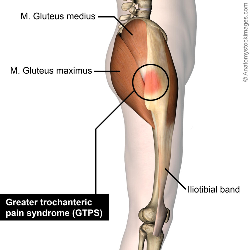
A bursa that sometimes causes problems in the hip is sandwiched between the bump on the outer hip (the greater trochanter) and the muscles and tendons that cross over the bump. This anatomical atlas was especially designed for a specific public (radiologists, surgeons, rheumatologists and physicians specializing in musculoskeletal imaging). A radiograph is not as helpful in diagnosing trochanteric bursitis as soft tissues and muscles are not visible to any degree(15). Microscopic anatomy of skeletal muscle. Leave a reply cancel reply. It is a flat, triangular muscle on the anterior wall of the pelvis. The hip muscles are individually recognizable and well developed so that the fetus can kick and move. Anatomy 3d atlas allows you to study human anatomy in an easy and interactive way. 936 x 504 png 317 кб. Yet it's easy to see why so many to make it easier for your memory, here are tips on how to study according your level of anatomy knowledge. This muscle assists with the external rotation of the hip. The muscles of the neck can be divided into groups according to their location. Your email address will not be published.
Learning the anatomy of your hip will better enable you to pinpoint your pain and work with your doctor to keep it from limiting your life. Learn about hip muscles human anatomy with free interactive flashcards. The muscles and the bones are under the layer of subcutaneous fat. Attached to the bones of the skeletal system are about 700 named. Leave a reply cancel reply.
Image Of Some Of The Anterior Hip And Thigh Muscles Of The Right Leg Thigh Muscle Anatomy Muscle Anatomy Hip Anatomy from i.pinimg.com Muscle movements, types, and names. This anatomical atlas was especially designed for a specific public (radiologists, surgeons, rheumatologists and physicians specializing in musculoskeletal imaging). Anatomy of the muscular system. The muscular system consists of the skeletal muscles and their associated structures. Highly detailed 3d models, with textures up to 4k resolution, enable to examine the shape of each. The hip muscles encompass many muscles of the hip and thigh whose main function is to act on the thigh at the hip joint and stabilize the pelvis. The hip is a complicated mechanism and therefore hip pain can originate in many different parts of the joint. Now that you watched the video, you.
The muscles of the neck can be divided into groups according to their location.
Several muscles cross the front of the hip and create hip flexion, pulling the thigh and trunk toward each other, but probably the most important is the iliopsoas. The muscular system is responsible for the movement of the human body. In human anatomy, the muscles of the hip joint are those muscles that cause movement in the hip. Muscles of the hips and thighs | human anatomy and. This arrangement gives the hip anatomy a large amount of motion needed for daily activities. The hip joint is the articulation of the pelvis with the femur, which connects the axial skeleton with the lower extremity. These muscles constitute the anatomical classification known as the medial compartment of the thigh. We study anatomy at the practical anatomy class we study the human body. Your email address will not be published. Anatomical terms allow us to describe the body and body motions more precisely. If left unstretched, shortened hip flexors affect the position of the pelvis, which in turn affects the position and movement of the lower back. Anatomy, bony pelvis and lower limb, psoas major. This muscle assists with the external rotation of the hip.
If left unstretched, shortened hip flexors affect the position of the pelvis, which in turn affects the position and movement of the lower back. The hip bone, also known as the innominate bone, coxal bone or os coxae, is a large bone that sits in the pelvis. The different anatomical areas of the gluteal region: Anatomy 3d atlas allows you to study human anatomy in an easy and interactive way. Most modern anatomists define 17 of these muscles, although some additional muscles may sometimes be considered.
Psoas Major Part I Hip Flexor Or Lumbar Stabilizer from www.sportsinjurybulletin.com The hip muscles encompass many muscles of the hip and thigh whose main function is to act on the thigh at the hip joint and stabilize the pelvis. Highly detailed 3d models, with textures up to 4k resolution, enable to examine the shape of each. The hip joint is a ball and socket joint that is the point of articulation between the head of the femur and the acetabulum of the pelvis. Learning the anatomy of your hip will better enable you to pinpoint your pain and work with your doctor to keep it from limiting your life. Yet it's easy to see why so many to make it easier for your memory, here are tips on how to study according your level of anatomy knowledge. The hip's unique anatomy enables it to be both extremely strong and amazingly flexible, so it can bear weight and allow for a wide range of movement. Anatomical terms allow us to describe the body and body motions more precisely. Muscle movements, types, and names.
Now that you watched the video, you.
Attached to the bones of the skeletal system are about 700 named. Your email address will not be published. Pick which works for you and then. A bursa that sometimes causes problems in the hip is sandwiched between the bump on the outer hip (the greater trochanter) and the muscles and tendons that cross over the bump. The cavity of the acetabulum the external obturator muscle is short external rotator muscle of hip joint. A radiograph is not as helpful in diagnosing trochanteric bursitis as soft tissues and muscles are not visible to any degree(15). The main functions of the neck muscles are to permit movements of the neck or head and to provide structural support of the head. The muscular system consists of the skeletal muscles and their associated structures. In utero fetal hips lie typically in flexion, abduction and external rotation, with the left hip usually muscular anatomy. The hip is a complicated mechanism and therefore hip pain can originate in many different parts of the joint. Diarthrodial joint with its inherent stability dictated primarily by its osseous components/articulations. The hip joint is a ball and socket joint that is the point of articulation between the head of the femur and the acetabulum of the pelvis. Muscle movements, types, and names.